Topigs Norsvin has a highly detailed and efficient system for CT data collection. These data are extremely valuable for pig breeding, but it can also be useful for developing imaging diagnostics in human medicine.
Written by: Øyvind Nordbø, PhD, Researcher at Topigs Norsvin.
Pigs and humans have relatively similar physiology. Therefore, pigs are often used as animal models in surgical training and biomedical research. Unlike humans, pigs have relatively short lifespans, so with pigs we do not face the same medical challenges, such as potential radiation damage from CT scanning.
In this article, we recount how, by sharing our knowledge and innovative solutions, Topigs Norsvin is contributing to advancements in human medicine.
Øyvind Nordbø, PhD, Researcher at Topigs Norsvin

Large-Scale CT Scanning at Topigs Norsvin
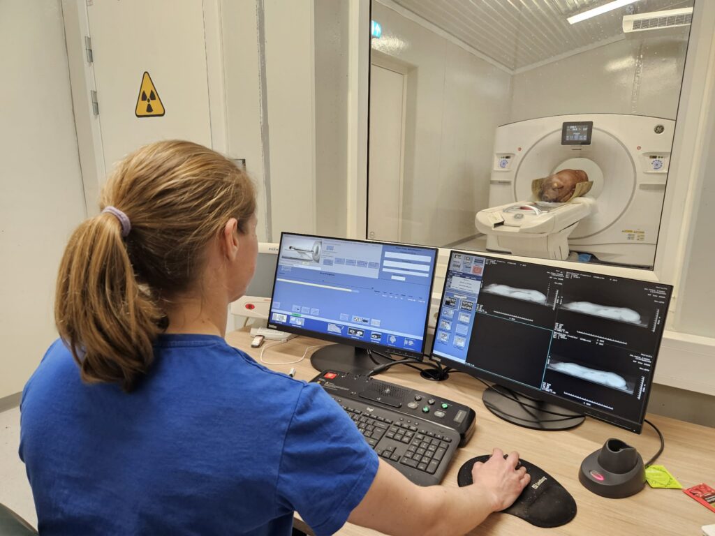
At Topigs Norsvin’s boar testing stations, Delta Norway and Delta Canada, several thousand pigs are CT scanned annually. In our breeding programs, CT scanning is used to obtain precise measurements of various carcass and health traits. Each pig is sedated before the scan, and the procedure has no noticeable side effects.
In the CT scanner, the subject is exposed to X-rays from different angles. Tissue types with different material densities, such as bone, muscle, and fat, absorb X-rays to varying degrees, and the grayscale in the CT image reflects the density of the scanned object. The scanner produces high-resolution 3D images, and Topigs Norsvin uses advanced computer models to extract precise measurements from the images of the scanned boars.
Challenges in Human Medicine
Establishing good medical datasets for humans faces two key obstacles. First, data sources must be completely anonymous to protect patient privacy. However, anonymization prevents researchers from linking multiple measurements from the same individual over time, making it difficult to analyze how genetics, environment, and lifestyle factors interact to influence health traits. This limitation does not exist in pig breeding, where complete individual records can be maintained throughout each animal’s life, allowing researchers to account for environmental factors when calculating genetic potential.
Second, radiation exposure is a significant concern. CT scanning uses X-ray radiation. For breeding boars, which live a maximum of two to three years, the radiation dose from a scan poses no health risk, as there is insufficient time for cumulative damage to develop. In human medicine, however, patients often undergo repeated scans over many years or decades, making radiation exposure a concern. A large, accumulated dose of X-rays increases the risk of cancer, so minimizing radiation exposure is a priority in human healthcare.
High-Quality Imaging with Reduced Radiation
To reduce long-term risks from CT scanning, the radiation dose can be lowered, but this compromises image quality. One possible solution is to train artificial intelligence (AI) to enhance images so that those taken with low radiation doses match the quality of normal-dose images. Training such AI models requires large amounts of data, which is difficult to collect because it would expose human test subjects to unnecessary radiation.
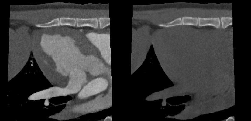
In collaboration with The Norwegian University of Science and Technology (NTNU) and Sorbonne University in Paris, Topigs Norsvin has performed repeated CT scans at normal, low (25%), and ultra-low (5%) radiation doses on 50 pigs (see Figure 1). These repeated measurements add only a few extra minutes on the CT table for each pig, making the additional cost relatively low . In return, Topigs Norsvin receives detailed annotated data of internal organs from its research partners, which can improve and expand the company’s AI models to provide a more detailed representation of pig anatomy.

Developing Cardiac CT Without Contrast
CT images work well for distinguishing tissues of different densities. However, blood and muscle have very similar densities, so contrast agents are often added to the blood for cardiac imaging. Contrast agents typically contain iodine, making the blood appear whiter than the heart muscle. Using contrast agents is not always desirable. Heart failure is often associated with kidney failure, and contrast agents can be harmful to people with poor kidney function. For such patients, Magnetic Resonance Imaging (MRI) is often used instead. However, MRI is much more expensive and time-consuming than CT, creating bottlenecks in hospitals.
An alternative to MRI and contrast-based cardiac CT is to develop AI that can differentiate blood and heart tissue in CT images obtained without contrast agents. At Topigs Norsvin, researchers have been working on a similar challenge through the project “With Heart for the Pig.” While contrast agents pose no health risk for pigs, they are not approved for use in food production.
Together with partners at The Norwegian University of Life Sciences (NMBU), the company has collected substantial cardiac CT data from pigs to develop novel traits relevant for cardiac function. Each pig was scanned twice: first without contrast, then a few seconds later with contrast. This dual-scanning approach creates a reference dataset that distinguishes muscle tissue from blood, enabling AI models to be trained on non-contrast CT images.
Open Data Initiative
These reference datasets are unique and have significant potential value for developing AI-based imaging diagnostics in human medicine. Recognizing this broader impact, Topigs Norsvin has decided to share them with various AI research communities.
So far, the company has provided access to this data for researchers at NTNU and Sorbonne University in Paris. Topigs Norsvin is now preparing to make portions of the dataset publicly available to researchers worldwide. By bridging the gap between agricultural research and medical innovation, this initiative demonstrates how data from livestock breeding can contribute to solving critical challenges in human healthcare.